3D PRINTED REPLICA OF THE CORROSION CAST DEMONSTRATING THE INTERNAL VASCULATURE OF THE HUMAN KIDNEY
Skrzat J 1, Walecki P 2, Tarasiuk J 3, Wroński S 3, Proniewska K 2
1 Jagiellonian University Medical College, Department of Anatomy,Cracow, Poland
2 Jagiellonian University Medical College, Department of Bioinformatics and Telemedicine, Cracow, Poland
3 AGH University of Science and Technology, Department of Condensed Matter Physics, Cracow, Poland
The most difficult for accurate 3D modelling seems to be intra-organ vascular network. The kidney is a good example of an organ which vascular pattern is simply in terms of the fractal geometry, however enough complicated for rapid prototyping to produce 3D models including all anatomical details.
The aim of this study is was to demonstrate possibilities of the 3D printing technology for fabrication replicas of the corrosion casts specimens demonstrating intra-renal vasculature.
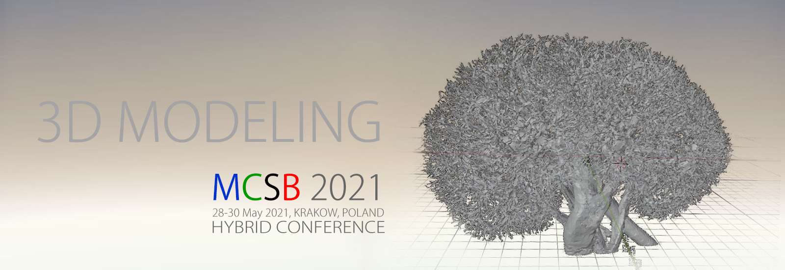
To prepare 3D models of the renal vasculature selected corrosion casts of the human kidney were scanned using a Nanotom 180N device produced by GE Sensing & Inspection Technologies Phoenix X-ray Gmbh. The spatial resolution of the reconstructed object was 60 µm. The reconstruction of scanned samples was done with the aid of proprietary GE software datosX ver. 2.1.0 using the Feldkamp algorithm for cone beam X-ray CT [3]. The post-reconstruction data treatment was performed by means of the VGStudio Max 2.1
Further, the MeshLab software was used for final preparing the printable 3D mesh model.
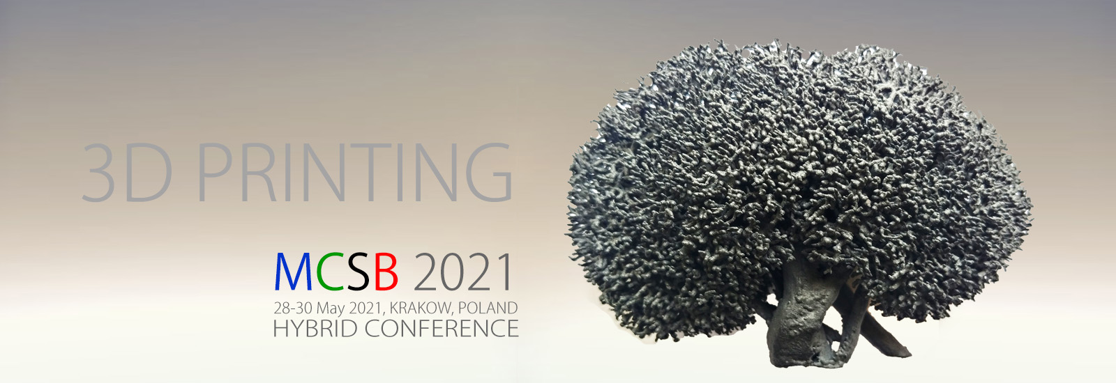
For materializing virtual mesh models of the renal vasculature we used desktop 3D printer Nobel 1.0 A. This 3D printing technology is based on stereolithography (SLA). The 3D printed models were created with photopolymer resin which solidified after exposed to the ultraviolet laser light, however remains flexible and mimics elasticity of the blood vessels. Alternatively, we also used powder 3D printer for manufacturing the same models. We used the Lisa 3D printer by Sinterit, which works in SLS (Selective Laser Sintering) technology.
3D printed models manufactured by means of the SLA 3D printer Nobel 1.0 replicated only main divisions of the renal vascular tree. Much better fidelity of anatomical details between 3D printed models and original corrosion cast specimens was attained using powder-based SLS 3D printing technology. Thus, obtained 3D models revealed correctly anatomical details of the vascular tree but we found minor artefacts being the oval endings of the terminal vascular branches, instead of the sharply pointed segments.
3D printed models of the renal vasculature system show high level of accuracy of subsequent divisions both in arterial and venous tree. Thereby, such models may serve for demonstrating normal vascular anatomy of the kidney, and the potential morphological anomalies of the intrarenal vasculature. Hence, the 3D printed models of the renal vasculature can be used for the medical students educational and for the surgeons practicing at the field of urology.


